Thursday, December 30, 2010

Sonography of renal sinus lipomatosis

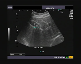
This middle aged patient presented with non specific complaints and we did a routine abdominal ultrasonography on him. This is what we found on ultrasound imaging of the kidneys:


Both kidneys showed echogenic matter within the region of the renal sinuses- and this was more prominent in the left kidney. The large echogenic mass like appearance in the lower part of the left renal sinus prompted us to think that this could be angiomyolipoma, one of the more common entities in the kidney. However, further imaging showed that this hyperechoic deposit was almost totally within the confines of the renal sinuses. Besides the lesions were bilateral.
 See ultrasound video clips below:
 
Further observe the vessels within the renal sinus of the left kidney in this color Doppler ultrasound video clip below:
 
The most obvious diagnosis based on these ultrasound images and video clips is renal sinus lipomatosis.
The uniformly homogenous appearance of the fatty tissue in the renal sinus suggests lipomatosis rather than any more ominous masses/ malignancies of the renal sinus. These ultrasound findings are suggestive of a grade -1 renal sinus lipomatosis of both kidneys.
References: http://www.jultrasoundmed.org/cgi/reprint/21/9/993 (free excellent article with images)
For more ultrasound/ color Doppler images of this case see:

Saturday, December 11, 2010

Subependymal cysts:

This neonate has a relatively large subependymal cyst close to the caudato-thalamic groove (ie the groove between the caudate nucleus and thalamus) of the fetal brain. Ultrasound videos taken in both the sagittal and coronal sections help confirm this diagnosis. The ultrasound video clip below shows a coronal section of the neonatal brain with the lesion displayed well.
 
The ultrasound video clip below shows a sagittal section through the neonatal brain displaying the subependymal cyst. The video clip pans from the lateral to the medial aspect of the neonatal brain, with focus on the subependymal cyst.


Still ultrasound images of this same neonatal brain are shown below: (see coronal images below):


This supendymal cyst is almost 7 to 8 mm. in dimension and needs careful follow up ultrasound imaging.
Parasagittal section image below shows another view of the subependymal cyst:


Visit:
http://www.ultrasound-images.com/pediatric.htm#Another_complication_of_subependymal_hemorrhage-_subependymal_cysts for more information and images of similar cases.

Saturday, December 4, 2010

Blunt trauma to testes with rupture:

This 25 yr. old male came to me with a history of severe blunt injury to the left scrotum. On inquiring further, he reported that the part of a heavy machine hit him squarely without causing any bleeding. However, the left scrotum was moderately swollen and markedly painful.
Ultrasound imaging of the scrotum revealed (see ultrasound video clip below):
 Observe the large hypoechoic (dark) inhomogenous area in the left testis. This is a typical appearance of an intratesticular hematoma. But is that all? The right half of the video shows partial rupture of the outer covering of the left testis (tunica albuginea). Do we see anything more? There is some spillage of the lateral part of the testicular parenchyma outside the confines of the testicular capsule.
See some more color Doppler ultrasound videos of this testicular trauma- below:
Observe normal vascularity (blood flow) of the main testicular tissue (in the above long section of the left testis). This is a good sign and shows that the injured left testis is viable and does not need to be removed surgically. However, the lower pole of the left testis does show vessels entering the tissue that has spilled out of the capsule of the testis. This confirms that seminiferous tubules from the testis have been pushed out of the rupture site (of the left testicular fibrous capsule). In addition, there is a moderately large hemorrhagic fluid collection in the left scrotum (left hematocele).
Visit this link to read more on this topic:
http://www.ultrasound-images.com/acute-scrotum.htm#Testicular_trauma_%28rupture_of_testes_or_fracture_testes%29:_
We have one more color Doppler ultrasound video (this time a transverse section of the left testis):
 
Observe the linear strip of tissue hanging (or floating) within the hematocele in the left scrotum. This could be left testicular tissue with part of the body of the left epididymis.
Here are the ultrasound/ color Doppler images of this case of testicular trauma:


This last image is a transverse section section of the testis showing the site of rupture of the tunica albuginea, using Power Doppler imaging. The intratesticular hematoma is also seen.

Tuesday, November 23, 2010

A small vascular lesion behind the thyroid

This color Doppler ultrasound image of the left lobe of the thyroid raised more queries than answers.
The lesion only 4 mm. in size. But located in a typical location in posterior aspect of the upper pole of left lobe of thyroid.


Markedly vascular lesions in this location are usually parathyroid adenomas. The gray scale ultrasound image suggests a solid lesion, thus suggestive of an adenoma of the parathyroid gland.


Such a highly vascular lesion can also suggest a possibility of an Arterio-venous malformation of the thyroid or the parathyroid. This is where this color Doppler ultrasound video might help (see below): this video shows a transverse section through the left lobe.


This color Doppler video clip below shows a long section of the left lobe of thyroid. The lesion is seen posteriorly.


See: http://www.ultrasound-images.com/thyroid.htm for more.
Specifically about parathyroid adenomas- visit:
http://www.ultrasound-images.com/thyroid.htm#Parathyroid_adenoma

Wednesday, November 17, 2010

Pedunculated cervical polyp- Ultrasound videos:

Polyps (small outgrowths) are known to occur in the cervical canal (the lumen of the lower third of the uterus), anytime from puberty to menopause. However, they are rare in menopausal women and even rarer in puberty. They are common in women over the age of 20 years and who have had children.
What is the cause of cervical polyps? It is believed that cervical polyps are the result of long term insult or inflammation of the lining (mucosa) of the cervical canal. Also, changes in estrogen levels can also cause polypoid growths to form in the cervical canal (endo-cervix).
What are the main symptoms of a polyp in the cervix? The common presenting symptoms are pain in the lower pelvis, vagina, discharge from the vagina- mucous or blood, irregular and painful menses etc.
What are the types of cervical polyps? The common types of polyps of the cervix are the sessile polyps (which have no stalk are seen as small projections from the endo-cervical lining). The rarer type is the pedunculated polyp of the cervix. This type of pedunculated cervical polyp has a long stalk connecting the growth to the lining  of the cervix. It may project into the vagina, and be confused with a endometrial polyp (a polyp of the body of the uterus) or small fibroid.
One the methods of diagnosing a cervical polyp is by ultrasound and color Doppler imaging of the uterus, best seen on transvaginal ultrasound (inserting the ultrasound probe via the vagina).
Present case (pedunculated polyp of the cervix):
This woman is an unusual case in that she is in the perimenopausal age (47 years) and has severe pain  in the lower pelvis with irregular menses.
This is what we saw on transvaginal ultrasound imaging (see sagittal section ultrasound video clip below):
 There is an elongated hyperechoic (bright) mass within the cervical canal (arrows).
 Power Doppler ultrasound video shows large feeder blood vessels supplying the mass (growth). This is a typical appearance. (See sagittal section Power Doppler video clip above).
Color Doppler ultrasound video clip (below) further confirms the highly vascular nature of the growth in the cervical lumen. It is elongated and fills the length of the cervix.
We performed a transverse section ultrasound imaging of the cervix, and this is what we saw (see the color Doppler video clip below):

The feeder vessels are seen entering and coursing through the entire polyp within the cervix.
Can such growths of the cervical lumen be cancerous? Generally, cervical polyps are benign (almost 99 % of polyps of the cervix are non cancerous). However, the highly vascular nature of the growth in this case, warrants further examination, at least a PAP smear and a study of the specimen after surgery is also advisable. 
 For further reading on this topic see this article on: Ultrasound imaging of Cervical polyps
For more ultrasound images of this case and more on imaging of the uterus, see:
http://www.ultrasound-images.com/uterus.htm

Tuesday, November 16, 2010

Mucosal edema of bladder following recent passage of calculus:

This patient underwent ultrasound imaging to rule out urinary stones. This is what we found in the urinary bladder:
The urinary bladder mucosa shows marked edema around the region of the left ureteric orifice (opening of left ureter into the urinary bladder). So could this be a malignancy or polyp of the bladder mucosa? Fortunately this patient gave a history of passing a calculus (stone) the same day. This suggests that the "mass" appearance in the left wall of the bladder is mucosal edema (mucosa is the inner lining of the urinary bladder), due to the stone being lodged in the left vesico-ureteric junction (distal end of left ureter).
Here are the ultrasound images of the same case:
The lesion is seen clearly. But the appearance is very similar to that of a bladder malignancy.
Color Doppler ultrasound image shows no significant vascularity (blood flow) within the mass (lump) (arrows). This again suggests a more benign nature of the mass.
The kidneys show small echogenic foci (one in each kidney) suggestive of renal calculi, with no backpressure  changes.
For more bladder related diseases and ultrasound study of these cases see:
http://www.ultrasound-images.com/urinary-bladder.htm

Thursday, November 11, 2010

Multiple Nabothian cysts of the cervix

 
This middle aged woman presented with irregular menses. Transvaginal ultrasound (see video clip above) shows multiple cysts of the cervix (transverse section of the cervix). This is the typical appearance of Nabothian cysts of the cervix. Nabothian cysts are also called mucinous retention cysts or Nabothian follicles of the cervix and are usually of little clinical significance unless complicated by infection or if they become unusually large. Most such cysts usually disappear without medical treatment. 
Here is a sagittal section ultrasound video of the same cervix (transvaginal):


As stated above, these Nabothian cysts of the cervix do not appear ominous in any way. However, the cause of this patient's complaints appear to be due to (see ultrasound images below):


The endometrium (the inner lining of the uterus) is seen to be markedly thickened (at 13 mm.). On color Doppler imaging of the uterus (both transabdominal and transvaginal), the endometrium does not show any vascular areas or increased blood flow within any part of it. This kind of thickening of the endometrium is called endometrial hyperplasia and needs further investigation. A pap smear of the cervical fluid showed some degree of squamous metaplasia. This patient needs to be monitored and might need treatment for the hyperplastic changes in the endometrium. 
For more ultrasound and color Doppler images of uterine pathology see:

Wednesday, November 10, 2010

Unusual bilateral ectopic fused kidneys:

The left and right kidneys appear to be ectopic with some degree of fusion of both kidneys, anterior to the mid line- the right kidney being located somewhere just below the liver and anterior to the spine; the left kidney located just behind the urinary bladder.
Have a look at this ultrasound video clip, panning from the bladder to the liver showing both kidneys, in the sagittal and oblique sagittal planes.


Some more ultrasound videos of the bilateral ectopic kidneys follow....
The left kidney is seen below- posterior to the urinary bladder in this B-mode ultrasound video (gray scale):


Have a look at the ectopic right kidney, located anterior to the spine, in this right parasagittal section ultrasound video clip:
Note the empty right renal fossa with the right kidney in an abnormal position.
Now the transverse section through the right kidney- see below:


References: http://www.springerlink.com/content/92207432001l2538/
http://www.ultrasound-images.com/kidneys.htm
S-shaped fusion of kidneys
Crossed renal ectopia (good article)
Pictorial display of various kidney fusion anomalies 
It is possible that the kidneys seen above may a variant of S-shaped or L-shaped fusion anomaly of the kidneys.

Friday, November 5, 2010

Normal Gallbladder variants- Phrygian cap:

The Phrygian cap shape of the gallbladder (Gall Bladder) is a known variant. See ultrasound image below:


The gallbladder shows a septate appearance in this image. But is this really a septate gallbladder? See ultrasound video clip below:
Still not sure? See the ultrasound video clip below:
 
The commonest cause for the"septate" appearance of the gallbladder is the Phrygian cap anatomical variant. For more on this topic see: http://www.ultrasound-images.com/gall-bladder.htm  The best method to demonstrate this variant of the GB is to unfold the gallbladder, by angling the probe to the length of the gallbladder- this displays the "cap" formed by the GB fundus. (See ultrasound picture below)-


The picture above shows the "unfolded" view of the gallbladder, with the Phrygian cap (arrow) clearly.

Thursday, November 4, 2010

Bowel mass- sigmoid carcinoma:

This middle aged male adult patient has a mass just above the urinary bladder. Ultrasound video clip of the mass is seen in sagittal section:
The mass shows an echogenic central area with hypoechoic, surrounding soft tissue area. This is called the "target sign" and is typical of colonic malignancy.
 Yet another sagittal ultrasound video clip shows the mass with the thickened sigmoid colon walls. Also note the absence of normal layered appearance of the normal large bowel.
This color Doppler ultrasound video shows the sigmoid colon mass to be poorly vascular. The mass has all the appearances of a sigmoid carcinoma.
 The close proximity of the mass to the urinary bladder (upper pelvis) and target sign are an indication of the mass originating from the sigmoid colon. The liver appeared normal. Close follow up and CT scan imaging are needed to rule out lymph node spread.
See more ultrasound images and description of this case at:
http://www.ultrasound-images.com/colon.htm
http://emedicine.medscape.com/article/367061-imaging

Tuesday, November 2, 2010

Double J -ureteric stent (ureteral stent):


This longitudinal section ultrasound video of the right kidney shows persistent mild hydronephrosis, despite the presence of the double J stent (DJ stent or ureteral stent) in the right ureter.
 

 The above ultrasound video shows the transverse section of the hydronephrotic right kidney with the upper end of the ureteral stent in the distended renal pelvis.
Reference: http://en.wikipedia.org/wiki/Ureteric_stent 
See the following link for article and images of ureteral stent:
See: http://cochinblogs.blogspot.com/2010/04/ultrasound-imaging-of-ureteral-stent.html 

Focal epididymitis:

This color Doppler ultrasound video shows a long section of the right testis- observe the lower pole.


The right tail of epididymis definitely appears inflamed. Now observe this transverse section color Doppler video clip through the right testis panning from the upper to the lower poles.


 There is a small right hydrocele which outlines the head and body of the right epididymis. The diagnosis in this case is focal right epididymitis involving primarily the tail.
Here is the still ultrasound image of the affected right testis:
And the color Doppler image of the same case: (right testis and epididymis):


And the still ultrasound image of the normal left testis (above).
For more on this topic see: http://www.ultrasound-images.com/scrotal-infections.htm

Saturday, October 30, 2010

Tuberculous epididymitis/ epididymo-orchitis:

This middle aged male patient presented with a mass in the left scrotum which was mildly painful. This ultrasound video shows a transverse section through the left scrotum, with the probe panning from the lower to upper poles of the left testis.
 
What do we see? Note the small hydrocele around the large, nodular hypoechoic mass in the lower pole of left testis. The mass originates in the tail of the left epididymis, and extends into the lower tip of the left testis.  In addition note the thickened scrotal wall. 
Have a look now, at the color Doppler and Power Doppler ultrasound videos of the left scrotum.


The above video clip is a color Doppler sonography study which shows marked vascularity of the "mass" in the tail of left epididymis. Note the thickened body of the left epididymis as we pan upwards.
What does this suggest? Clearly the mass is primarily a lesion involving the left epidiymis from body to the tail and the small hydrocele all point to an inflammatory pathology. The commonest cause of such an appearance is tuberculous epididymitis with orchitis (involvement of the adjacent left testis). The 2nd diagnostic possibility in such a case is pyogenic/ non tuberculous orchitis- in non tuberculous orchitis or non tuberculous epididymitis, the epididymis would be homogenously enlarged, compared to the inhomogenous mass that is seen in this case. See: Tuberculous epididymo- orchitis article(AJR)  
  This sagittal to parasagittal section Power Doppler ultrasound video clip of the left testis further confirms the findings mentioned above.
The last video- a zoomed in view of the mass:

Thursday, October 28, 2010

Chronic pyelonephritis- atrophic kidney:

This adult male patient had recurrent calculi in the right kidney. Ultrasound video clip (long axis view) of the right kidney shows:
 
Note the small right kidney with irregular contour suggesting chronic infection/ pyelonephritis.
  
Color Doppler video clip of right kidney shows poor vascularity in the right kidney, again confirming atrophic changes and poor functionality in this kidney.

 
The normal left kidney is shown for comparison in this color Doppler ultrasound video clip. There is a certain degree of compensatory hypertrophy in this kidney.

Tuesday, October 26, 2010

Pelvic kidney- ultrasound video study

This young adult female patient presented with right hypochondrial pain. We conducted a sonography of the gall bladder and found:
 
There were at least 3 calculi in the gall bladder, each of 7 to 9 mm. (see ultrasound video clip above) and the image below:
But a scan of the right kidney showed an empty right renal fossa:
So are we looking at a case of agenesis (absence) of the right kidney?
We searched further below and found this reniform structure near the urinary bladder.


Here are the still ultrasound images of what we saw....


At 6.6 x 2.5 cms., this kidney shaped structure looked rather small. But ultrasound and color Doppler appearances convinced us that this is indeed an ectopic right kidney located in the right hemipelvis.


 The above color Doppler ultrasound video clip further confirms our findings- this is indeed a pelvic right kidney. See: http://www.ultrasound-images.com/kidneys.htm for more on this topic.
Conclusion: the right hypochondrial pain was due to cholelithiasis with the pelvic (ectopic) right kidney an incidental finding.

Wednesday, October 20, 2010

Normal third trimester fetal anatomy -ultrasound videos: the abdomen:

This was a normal 32 week old fetus. One of the best ways to visualize the fetus is a cross sectional anatomy. But since one plane alone is not sufficient, we tried to take sections along various oblique planes.
The first ultrasound video is that of the right dome of fetal diaphragm:
The fetal diaphragm is seen as a hypoechoic curved line between the liver and the right lung. Observe the movements of the diaphragm with fetal respiratory attempts.
(abbreviations: LG= fetal right lung; H= fetal heart; LIV= fetal liver)
 Here is a still ultrasound image of the right dome of diaphragm.

Fetal Liver and Gall bladder:
Perhaps the easiest and most prominent structure in the fetal abdomen is the liver. This is seen as a large echogenic structure occupying the right hypochondrium of the fetus with a number of vessels passing through it. The fetal gall bladder is seen within this structure (the liver) as an anechoic pear shaped, elongated structure. The gall bladder is best appreciated in color Doppler imaging.
Note in the above color Doppler ultrasound video, that the gall bladder shows no flow unlike the umbilical vein and other nearby vessels.
  
This still ultrasound picture shows the gall bladder through the fetal liver:
 And this color Doppler image helps differentiate the gall bladder from the vessels nearby:

The fetal stomach:
Visualization of the fetal stomach is important to rule out esophageal atresia, pyloric stenosis and duodenal atresia. The fetal stomach is seen as an anechoic bubble in the left upper quadrant of the fetal abdomen.


This ultrasound video clip shows the normal fetal stomach (arrow) to the left of the spine in the left hypochondrium. Care must be taken to determine if the stomach is indeed on the left side of the abdomen and thus rule out situs anomalies (situs inversus etc).
Ultrasound image showing the normal fetal stomach- the dark "bubble" - to the left of the liver:
 
Fetal Kidneys:

 
The fetal kidneys must be visualized and measured. Aplasia, hypoplasia or absence of one or both kidneys must be ruled out. Also, the sonographer/ radiologist must look for renal masses, cysts and hydronephrosis. Pelviureteric junction obstruction and posterior urethral valve in the fetus are common findings and important causes of fetal hydronpehrosis. The above video clip shows the normal left fetal kidney, next to the spine.
Still image of the normal left kidney in fetus:


The right fetal kidney is seen in this ultrasound video clip.
Still ultrasound image of normal right kidney: (long axis view)-

 
See this 2nd video clip of the normal right kidney. Note the hypoechoic triangles within the kidneys- these are normal renal pyramids (part of the renal medulla). Observe how the fetal kidneys move with fetal respiratory movements. 
Both kidneys must be visualized in both long and transverse axes: this transverse section ultrasound image shows both fetal kidneys-
Fetal adrenal (suprarenal) glands:


The fetal adrenals (suprarenal glands) must be carefully visualized to rule out abnormalities including masses (neuroblastoma) and cysts. The suprarenal glands are seen as triangular caps above the upper pole of each kidney. They have an echogenic central part with a hypoechoic layer outside. The above video clip shows a transverse section through the right suprarenal gland.
A still image of the fetal right adrenal gland is seen below (this is an oblique section through the gland):
One more ultrasound image of the right suprarenal gland is shown here- ( the image below is a coronal section of the right kidney and the right suprarenal gland)


This is a longitudinal section through the right kidney and right adrenal gland. 
Visit: http://www.ultrasound-images.com/fetal-abdomen.htm for more images and abnormal conditions/ images about this topic.
Umbilical vein (intra fetal part):


The umbilical vein is seen coursing through the liver (color Doppler ultrasound video clip-at right). This vein carries oxygenated blood from the placenta to the fetus.
The fetal abdominal aorta and the iliac arteries:
The normal abdominal aorta is seen in this coronal section ultrasound image, dividing near the pelvis into the common iliac arteries (arrows).

Tuesday, October 19, 2010

Unusual case of right flank pain:

This elderly male patient had right flank pain. We suspected a renal or ureteric calculus.
The ultrasound video above shows a "lump" in the right kidney in the middle third of the renal cortex. Is this a renal mass? The sonography of this case speaks in riddles- there is a vague ill-defined mass like area, but there is also a definite, but mild dilatation of the pelvicalyces. This is a case of right ureteric calculus with Bertin's column involving the right renal cortex.
Here is a still image of the same case:
Arrows point to the lesion (a normal variant). Note the dilated pelvicalyces.
The ultrasound video clip above shows the left kidney of the same patient- an obvious small cortical cyst is seen. There were no symptoms related to the left flank.
And here is the still image showing both kidneys:


See:

Monday, October 18, 2010

Benign prostatic hypertrophy- TRUS ultrasound videos

This was a rather large hypertrophy of the prostate in an elderly male patient. Prostates can get bigger than this. This ultrasound video demo of benign hypertrophy of the prostate was obtained via the transrectal route. Often, large prostates can be difficult to image in transrectal ultrasound studies due to the sheer size of the organ. Hence it is prudent to first image huge prostates via the transabdominal route, with a full bladder before trying the endocavity method. I always prefer to image prostates first in the oblique coronal plane from apex to base (as in this ultrasound video).
This is a still image of the prostate in the same patient.
See: http://www.ultrasound-images.com/prostate.htm for more images and discussion of prostate pathology.

Case-2:
This color Doppler ultrasound video shows a large prostate (benign enlargement) with marked vascularity on color Doppler imaging:
This amount of hyperemia of the prostate is not normal and can't be explained by benign enlargement. The diagnosis here is benign hypertrophy with prostatitis; something that can add insult to injury with marked exacerbation of the urinary complaints.

Saturday, October 16, 2010

Transrectal ultrasound videos- seminal vesicles and vas deferens:

This video shows the left vas deferens as it merges with the left seminal vesicle to form the ejaculatory duct:
                                                                        See: http://www.ultrasound-images.com/prostate.htm for more on this topic. This includes normal sonographic anatomy during TRUS (transrectal ultrasound) imaging for the prostate, seminal vesicles, vas deferens and the ejaculatory ducts. Of these the prostate is the largest and most obvious structure seen. In the oblique coronal plane, whilst angling the transrectal/ endocavity ultrasound probe upwards, one sees the 2 seminal vesicles seen resembling a bow tie. Further tilting the probe upwards reveals the vas deferens - 2 narrow tubes joining the seminal vesicles. The ejaculatory ducts are often difficult to see unless dilated due to pathology.                   

                                                                            
The above endocavity (transrectal) ultrasound video shows the right vas deferens merging similarly with the right seminal vesicle.
The ultrasound video clip below visualizes the seminal vesicles and vas of both sides:

                                  
And finally, imaging the prostate from the apex (or lower tip) and sweeping upwards towards the seminal vesicles. This way one sees almost every region of interest in TRUS study.
                                                                 Another important view is the sagittal section of the prostate which shows the posterior urethra or prostatic urethra as it is known. This view is also important to show potential calculi within the prostatic urethra, as also prostatic cysts in relation to it. Pressure on this part of the urethra can cause mild to severe urinary symptoms, usually caused by either of the pathologies mentioned above.

Friday, October 15, 2010

An Unusual cortical cyst of kidney- ultrasound video:

       
This cortical cyst is a rather unusual location in the left kidney- it occupies a central part (the middle third) of the kidney extending from the outer cortex to the renal pelvis. This appearance can mimic a calyceal diverticulum or caliectasis.
 The ultrasound image above shows the renal cortical cyst in transverse and longitudinal axes. Read more on this at: http://www.ultrasound-images.com/kidneys.htm 
And this is what a typical cortical cyst of the kidney looks like:
There is mild pelvicalyceal dilatation of the left kidney, due to pressure effect from the large left renal cortical cyst in the upper pole. I have seen renal cortical cysts from 0.5 cms. to 8 cms. in size. These cysts need to be monitored closely for signs of rupture, infection and malignancy.
  
 This color Doppler video clip of the same patient shows vessels curving around the rim of the cortical cyst of the left kidney.
Case-2:
A typical cortical cyst of the left kidney ( a very large one):

An ultrasound video clip of this large cyst in left kidney- it measures 6 to 7 cms. in diameter!
It is not unusual for renal cortical cysts to reach this size. Fortunately, there is no evidence of infection, hemorrhage or septation within this cystic lesion. But it needs to be periodically monitored for complications.

Wednesday, October 13, 2010

Complex kidney cysts (an ultrasound video study):

                                                                          
Case-1:Sagittal section ultrasound video clip showing the complex nature of the right renal cyst. This cyst is small and septate (fine septations). Note the presence of small calcifications in the lower part of the cyst.
Same case as above- another ultrasound video clip:
                                                                            
This cyst was seen in a middle aged male patient and was an incidental finding. CT scan reports suggest this is a Bosniak category 2 complex renal cyst. Indeed the ultrasound findings did not change over a period of time, nor did the cyst increase in size. This complex renal cyst measures 1.7 cms. in size.                                          
Case-2:
                                                                        The above ultrasound video clip shows a larger complex cyst in the left kidney of an elderly male patient. Again, the cyst might fit in with a Bosniak category-2 complex renal cyst. This renal cyst shows fine (but thicker than those in case-1 above) membranous partitions and a few calcifications along the cyst wall.                                                                                          
                                                                       This 2nd renal ultrasound video clip shows that there is a 2nd smaller cyst adjacent to the larger cyst. This one also shows few calcific lesions along the wall. These appearances suggest a benign nature of these complex cysts of the left kidney.                                                                                                                                   
                                                                          
 The above color Doppler video clip of the left kidney (same case-2) shows no significant vessels along the walls of the cysts or in the septae within them, again confirming the benign nature of these lesions.                    Read more at: http://www.kidneycancerinstitute.com/kidney-cyst.html
Also view ultrasound images of renal cysts and cystic diseases of kidneys at:
  http://www.ultrasound-images.com/kidneys.htm

Friday, October 8, 2010

A difficult mid-ureteric calculus:


This patient had right flank pain since a few months. But was not sure if he had a renal stone (calculus). The bladder ultrasound image appears normal.


The left kidney is normal. But not the right kidney- shows moderate hydronephrosis (back-pressure changes). The stone must be lower down somewhere....A difficult job to find this calculus, somewhere in the right ureter.. in a moderately obese patient.


Persistence and dogged determination to find the culprit, works. Sustained graded compression over the right flank, shows the dilated right ureter and the 17 mm. calculus in mid ureter.


Recorded an ultrasound video clip of the right mid ureteric calculus (arrows).
  See: http://www.ultrasound-images.com/ureteric-calculi.htm for more on this topic.

Wednesday, September 29, 2010

Popliteal vein thrombus- Color Doppler ultrasound video clip:

                                                                               This color Doppler video clip shows the thrombus (arrows), clearly, in transverse section of the vessel.
                                                                       And this- a Color Doppler video clip of the same vessel in longitudinal section.
See:  http://www.ultrasound-images.com/vascular.htm (for more on this case). On that page, you should find more color Doppler images of this case and details of how to image the veins of the lower limb. The interesting and indeed intriguing part of this case is the particulate movement within the thrombus- possibly fibrotic strands within it. This suggests that the thrombus is old or chronic, maybe a month or more old. 

Thursday, September 23, 2010

Cystic duct- ultrasound video:

                                                                             Color Doppler video clip showing the normal cystic duct as it emerges from the gall bladder to merge with the common bile duct. The portal vein is seen adjacent to the cystic duct.                                                       See: http://en.wikipedia.org/wiki/Cystic_duct  for more.                                                                           Also:   http://radiographics.rsna.org/content/21/1/3/F1.expansion                                                                
 The color Doppler image above shows the cystic duct clearly. PV= portal vein.

Wednesday, September 22, 2010

Color Doppler video- simple cyst of testis:

This male patient underwent sonography of the scrotum for left testicular pain. The color Doppler video clip of the left testis shows a simple cyst of the upper pole of left testis. In addition, the patient has a large, grade 3 varicocele of the left scrotum (see video below). The cause of the left scrotal pain is in all probability, the varicocele, the cystic lesion of the testis being only an incidental finding.
                                                                                                                                                                                                                                   
Here are the still images demonstrating the simple testicular cyst:


The above ultrasound image shows a transverse section through the upper pole.  
 This is a long section color Doppler image of left testis with the non vascular anechoic lesion seen in the upper pole, clearly a cyst.