This article is about ultrasound imaging of the fetal heart and in particular the 3 vessel view. After the four chamber view of the fetal heart, the most important view is the 3 vessel view and that too the imaging of the fetal ductus arteriosus. This vessel (the ductus arteriosus) is the direct continuation of the fetal pulmonary trunk and is part of the trifurcation of the pulmonary trunk into three main vessels- the right and left pulmonary arteries and the ductus ateriosus. It requires a lot of patience of image the fetal ductus ateriosus in its entirety and often is the exercise can be frustrating. I had a lot of trouble identifying the ductus arteriosus correctly and hence I have captured a good number of videos and dedicated many articles to this topic.
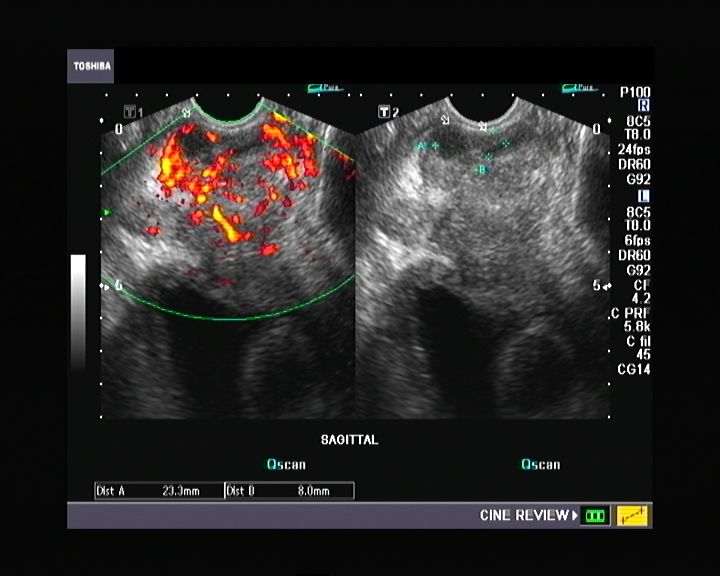
Here are some good fetal echo 3 vessel view images:

Ultrasound images of the ductus arteriosus and its relation to the arch aorta as they both meet in a V shape posteriorly:


ANTR= ANTERIOR
POSTR= POSTERIOR
DUCT= DUCTUS ARTERIOSUS
DESC AO= DESCENDING AORTA
PA= PULMONARY ARTERY
AA= ARCH OF AORTA
SVC= SUPERIOR VENA CAVA
The ultrasound video clip below shows the 3 vessels, namely the superior vena cava (on the right), the aorta (middle) and pulmonary artery (left). The sequence of the 3 vessels can be memorized from right to left by the word- SAP. The ductus is at the bottom (left) of the images and is seen meeting the arch of the aorta with which it forms a V shape. (see: http://www.ultrasound-images.com/normal-anatomy.htm )