Wednesday, December 25, 2019

Ultrasound-video-GB-calculi

A common finding in middle aged female patients- multiple gallbladder calculi.
This lady had no complaints or symptoms.
for more on gallbladder diseases:
https://www.ultrasound-images.com/gall-bladder/

Wednesday, December 11, 2019

Aortic'and-mitral-valves-calcification

Age related changes of aortic and mitral valves, in this case dense calcification. Also present are wall motion abnormalities of LAD territory. 

https://youtu.be/hWHbblJp-18

Sunday, November 24, 2019

encephalocele-ultrasound-video

This 2nd trimester fetus shows a large occipital encephalocele.
This encephalocele was huge at 3.5 cms.


Saturday, June 1, 2019

Huge-umbilical-hernia

This umbilical hernia protruded from the umbilicus and was 15 cms diameter.
Ultrasound video of the umbilical hernia shows peristaltic movement of small bowel loops within.

Tuesday, March 26, 2019

Atlas of neonatal ultrasound

My newest e-book Atlas of Neonatal Ultrasound is available on amazon Kindle.
see this link to download this e-book on your mobile, either android or iphone
Here is the link again: https://www.amazon.in/dp/B07M5W7NZ4
It is an e-book that's designed to give the reader, a sonologist, radiologist or sonographer a rapid review of the common cases one comes across during sonography of neonates. 
For those in USA and Canada it is at: https://www.amazon.com/dp/B07M5W7NZ4
Happy reading. 👸


Monday, December 3, 2018

Best echocardiography videos

I found these two videos to be very good for echocardiography basics:
I think this is East European or Russian
But narrative is in English.

https://youtu.be/7WNc2ND32Hk

Saturday, July 28, 2012

Some useful free video editing software

http://www.freemake.com/free_video_converter/ 
This is a very useful free software/ freeware to convert videos from avi, mpg, mpg4 etc. to various formats as well as for converting audio files. A must have. I've used it and there were no bugs detected.


Want to join video clips together in a couple of clicks? Free video joiner is the freeware that does this in a couple of clicks. Easy to use and a very simple interface:
http://www.freevideojoiner.com/

Zweistein is also a simple and free software that is a great video editor.
You can use its crop function to remove those identity details from video clips (remove the names of patients and other such ID details) from the borders of video clips.
See: http://www.thugsatbay.com/tab/?q=zweistein


Thursday, July 19, 2012

Ultrasound imaging of large epigastric hernia

Video clip showing ultrasound imaging of this large epigastric hernia in a middle aged man:


Ultrasound images show the large midline defect in the abdominal wall with hernial sac protruding through the epigastric defect:


See:
https://www.ultrasound-images.com/musculoskeletal/
 for more on this topic

Friday, July 13, 2012

Inguinoscrotal hernia -ultrasound imaging


The hernial sac is seen above the right testis in this ultrasound video of the right scrotum. A small amount of fluid is seen in the hernial sac. The video shows a few bowel loops with peristalsis within the Rt. inguinoscrotal hernia.
More ultrasound images of this case are shown below:
(Machine used is the Philips HD 15 system):


See more cases of inguinal hernia at: http://www.ultrasound-images.com/scrotum.htm#Indirect_inguinal_hernia_forming_large_Right_scrotal_swelling

Wednesday, July 11, 2012

Sonography of the fetal ductal arch


   A sagittal section of the fetal ductal arch (see the ultrasound video above) shows the typical hockey stick shape of the ductal arch as it emerges from the fetal right ventricle. Observe closely, that in sagittal section the fetal right ventricle is the chamber that is adjacent to and just behind the anterior chest wall (the sternum) of the fetus.
  The fetal aortic arch, on the contrary is an umbrella shaped structure and is seen to arise more posteriorly (see ultrasound image below).


Tuesday, April 3, 2012

Normal fetal echo quiz

Name the structures marked by the numbers 1,2 and 3 in this fetal echo of a 28 week fetus:
Still image is shown below:


The ultrasound video of this same case is shown below:


Hint: see: http://www.ultrasound-images.com/normal-anatomy.htm#Normal_3_vessel_view_in_a_28_week_fetus

Sunday, April 1, 2012

The ductus and 3 vessel view in 30 week fetus

Just some stunning ultrasound and color Doppler images and a video of the 3 vessel view during fetal echo in a 30 week fetus. In the color Doppler image below, the right pulmonary artery is seen crossing behind the ascending aorta (left half of image). The ductus arteriosus (red) is seen merging with the aortic arch (blue) to the left of the spine (right half of image).


Image below shows details of the 3 vessel view.


The video below shows the 3 vessel view again in this 30 week fetus. The visualization of the ductus may vary depending on imaging conditions.

Thursday, March 29, 2012

Fetal echocardiography basics- the ductus arteriosus:

This article is about ultrasound imaging of the fetal heart and in particular the 3 vessel view. After the four chamber view of the fetal heart, the most important view is the 3 vessel view and that too the imaging of the fetal ductus arteriosus. This vessel (the ductus arteriosus) is the direct continuation of the fetal pulmonary trunk and is part of the trifurcation of the pulmonary trunk into three main vessels- the right and left pulmonary arteries and the ductus ateriosus. It requires a lot of patience of image the fetal ductus ateriosus in its entirety and often is the exercise can be frustrating. I had a lot of trouble identifying the ductus arteriosus correctly and hence I have captured a good number of videos and dedicated many articles to this topic.
Here are some good fetal echo 3 vessel view images:


Ultrasound images of the ductus arteriosus and its relation to the arch aorta as they both meet in a V shape posteriorly:


ANTR= ANTERIOR
POSTR= POSTERIOR
DUCT= DUCTUS ARTERIOSUS
DESC AO= DESCENDING AORTA
PA= PULMONARY ARTERY
AA= ARCH OF AORTA
SVC= SUPERIOR VENA CAVA

The ultrasound video clip below shows the 3 vessels, namely the superior vena cava (on the right), the aorta (middle) and pulmonary artery (left). The sequence of the 3 vessels can be memorized from right to left by the word- SAP. The ductus is at the bottom (left) of the images and is seen meeting the arch of the aorta with which it forms a V shape. (see: http://www.ultrasound-images.com/normal-anatomy.htm  )

Thursday, March 22, 2012

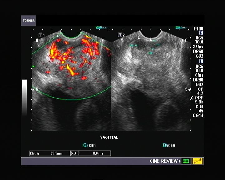
Prostate -transrectal ultrasound quiz

This patient has a nodular prostate on per rectal digital examination. What is your diagnosis based on these ultrasound images and video (TRUS scan) of the prostate:


Monday, January 2, 2012

Incompetent perforators and varicose veins: part-2

This is the same patient as in the previous blog. Here one of the perforators of the lower third of the right leg is seen between the right posterior tibial veins (paired) and the distended great saphenous vein.
To get this video, I passed the probe along the leg from below (ankle level) upwards, sweeping in the transverse plane until the perforator was seen clearly.
Color Doppler ultrasound Video 1:


Video-2: Another display of the same perforator:


The transverse axis of the great saphenous vein (GSV) is ideally suited to display the perforator emerging from the GSV and reaching deeper towards the posterior tibial veins (seen here as paired veins on either side of the posterior tibial artery).

Ultrasound video sapheno-femoral incompetence: part-1:


This patient of right lower limb varicose veins presented a strange display of the incompetent valves flailing helplessly in the to and fro flow of blood from and to the right great saphenous vein across the right saphenofemoral junction.
The spectral Doppler trace of this junction proves the point with moderately severe incompetence seen as reflux across the valve lasting almost 2 seconds. (see image below):


For more on this topic visit: http://www.ultrasound-images.com/vascular.htm#Chronic_venous_insufficiency_%28Incompetence_of_great_saphenous_vein%29

Saturday, December 17, 2011

Fine needle aspiration and biopsy of hemorrhagic thyroid cyst- case-2:

Ultrasound guided F\fine needle aspiration of fluid from a hemorrhagic thyroid cyst (see ultrasound video below):


The ultrasound images of the left lobe hemorrhagic cyst before and after aspiration of bloody thick fluid under ultrasound guidance:
The cyst measures 2.4 x 2.3 cms. before aspiration; observe the large amount of anechoic fluid within the large cyst in the left lobe. The right lobe appears normal. 


After aspiration the hemorrhagic cyst looks shrunken and collapsed; the cyst measures 1.9 x 2.2 cms.
 

The cyst looks more hyperechoic with loss of fluid content:
 
Further aspiration and the cyst collapses further. 
 

Thursday, November 24, 2011

FNAC of thyroid cyst:


This was a large cystic lesion of the right lobe of thyroid. Ultrasound guided fine needle aspiration of the cyst helped to drain the contents of the cyst as well as to ascertain the nature of the cyst contents. Ultrasound guided aspiration done here shows the needle tip descending into the cyst (seen as the bright mobile echogenic focus in the cyst lumen). The ultrasound probe (here) is placed perpendicular to the long axis of the needle- resulting in the needle tip being visualized. If the needle's long axis is parallel to the ultrasound beam (and the ultrasound probe), a linear echo of the needle can be visualized.
See: http://radiographics.rsna.org/content/28/7/1869.full 

Friday, November 11, 2011

Sonography quiz- What is your diagnosis?

This was a male patient with history of hematuria. What is your diagnosis? This ultrasound video shows a transabdominal scan in the first 2 parts of the video; the last part of the video shows a Transrectal ultrasound (TRUS) imaging of the bladder of the urinary bladder.


Answer: this is a freely mobile calculus in the urinary bladder. Seen rolling across the inner bladder surface on change in position. The TRUS imaging of the this case needed a lot of patience to capture this rather elusive urinary bladder stone.