Thursday, March 22, 2012

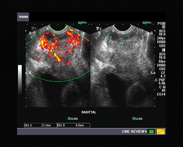
Prostate -transrectal ultrasound quiz

This patient has a nodular prostate on per rectal digital examination. What is your diagnosis based on these ultrasound images and video (TRUS scan) of the prostate:


Monday, January 2, 2012

Incompetent perforators and varicose veins: part-2

This is the same patient as in the previous blog. Here one of the perforators of the lower third of the right leg is seen between the right posterior tibial veins (paired) and the distended great saphenous vein.
To get this video, I passed the probe along the leg from below (ankle level) upwards, sweeping in the transverse plane until the perforator was seen clearly.
Color Doppler ultrasound Video 1:


Video-2: Another display of the same perforator:


The transverse axis of the great saphenous vein (GSV) is ideally suited to display the perforator emerging from the GSV and reaching deeper towards the posterior tibial veins (seen here as paired veins on either side of the posterior tibial artery).

Ultrasound video sapheno-femoral incompetence: part-1:


This patient of right lower limb varicose veins presented a strange display of the incompetent valves flailing helplessly in the to and fro flow of blood from and to the right great saphenous vein across the right saphenofemoral junction.
The spectral Doppler trace of this junction proves the point with moderately severe incompetence seen as reflux across the valve lasting almost 2 seconds. (see image below):


For more on this topic visit: http://www.ultrasound-images.com/vascular.htm#Chronic_venous_insufficiency_%28Incompetence_of_great_saphenous_vein%29

Saturday, December 17, 2011

Fine needle aspiration and biopsy of hemorrhagic thyroid cyst- case-2:

Ultrasound guided F\fine needle aspiration of fluid from a hemorrhagic thyroid cyst (see ultrasound video below):


The ultrasound images of the left lobe hemorrhagic cyst before and after aspiration of bloody thick fluid under ultrasound guidance:
The cyst measures 2.4 x 2.3 cms. before aspiration; observe the large amount of anechoic fluid within the large cyst in the left lobe. The right lobe appears normal. 


After aspiration the hemorrhagic cyst looks shrunken and collapsed; the cyst measures 1.9 x 2.2 cms.
 

The cyst looks more hyperechoic with loss of fluid content:
 
Further aspiration and the cyst collapses further. 
 

Thursday, November 24, 2011

FNAC of thyroid cyst:


This was a large cystic lesion of the right lobe of thyroid. Ultrasound guided fine needle aspiration of the cyst helped to drain the contents of the cyst as well as to ascertain the nature of the cyst contents. Ultrasound guided aspiration done here shows the needle tip descending into the cyst (seen as the bright mobile echogenic focus in the cyst lumen). The ultrasound probe (here) is placed perpendicular to the long axis of the needle- resulting in the needle tip being visualized. If the needle's long axis is parallel to the ultrasound beam (and the ultrasound probe), a linear echo of the needle can be visualized.
See: http://radiographics.rsna.org/content/28/7/1869.full 

Friday, November 11, 2011

Sonography quiz- What is your diagnosis?

This was a male patient with history of hematuria. What is your diagnosis? This ultrasound video shows a transabdominal scan in the first 2 parts of the video; the last part of the video shows a Transrectal ultrasound (TRUS) imaging of the bladder of the urinary bladder.


Answer: this is a freely mobile calculus in the urinary bladder. Seen rolling across the inner bladder surface on change in position. The TRUS imaging of the this case needed a lot of patience to capture this rather elusive urinary bladder stone.

Saturday, November 5, 2011

Ultrasound guided thyroid FNA biopsy:

FNA or fine needle aspiration of the thyroid is mainly used to distinguish benign from malignant thyroid nodules. However, it is also useful in the biopsy of diffuse enlargement of the thyroid as is seen in Hashimoto thyroiditis and diffuse infiltration of the thyroid in lymphomas and certain forms of metastases (colonic adenocarcinoma spreading to the thyroid). In the case of Hashimoto thyroiditis, FNA biopsy helps to distinguish Hashimoto thyroiditis from lymphomas and papillary carcinoma of the thyroid which may coexist with the primary disease.

Method: the aim of ultrasound guidance whilst performing the FNA biopsy is provide visualization of the needle tip and guide it to the exact location within the thyroid where the nodule is located or where there is maximum possibility of collecting diseased tissue. This short ultrasound video clip shows the needle tip (seen as an echogenic linear focus) moving to and fro within the left lobe of the thyroid. The aim of the FNA study in this case was to rule out lymphoma involving the thyroid:


References: http://radiographics.rsna.org/content/28/7/1869.full
(free article on Ultrasound guided FNA biopsy of the thyroid).